Bangyan Huang Wins Best Paper Award for Not-So-SIMPLE Work on PET Imaging
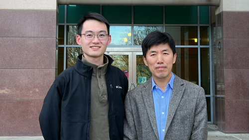
Bangyan Huang, a second-year doctoral student at the Department of Biomedical Engineering, was recently awarded first place in the 2022 Christopher J. Thompson Best Student Paper Award for his oral presentation “Statistical Image Reconstruction of Positron Lifetime via Time-Weighting (SIMPLE).” Huang received the award at the IEEE Medical Imaging Conference, which was held both online and in Milan, Italy in November 2022.
“Winning this award makes me motivated to do a lot more for this field,” he said. “I didn’t have a lot of hope because I gave the presentation remotely at 2 a.m., but actually I think it was a good thing because I presented slower, which allowed people to better absorb and understand this research that is very new in this field.”
The award recognizes outstanding student contributions and greater student participation as principal or sole authors of papers, as well as to acknowledge the importance of student contributions to the fields embraced by the Nuclear Science Symposium.
Although the acronym of Huang’s presentation is SIMPLE, the research is anything but. He and his faculty advisor, Professor Jinyi Qi, have developed a new concept for positron emission tomography (PET) imaging that uses positronium lifetime as new biomarkers for identifying abnormalities in tissue in PET scans. It is work that has specific applications in fighting cancer and Alzheimer’s disease.
Conventional PET scans use a radioactive drug, or a tracer, to show normal and abnormal metabolic activity in a body. Huang and Qi’s method instead uses positronium lifetime imaging—a biomarker that has never been visualized before.
Positronium is a system consisting of an electron and its antiparticle, a positron, bound together into an exotic atom. Positronium lifetime is what carries information about the tissue microenvironment where positrons are emitted. Previously, positronium lifetime hadn’t been used because of low sensitivity in detection and low spatial resolution of time-of-flight capabilities of PET scanners.
However, Qi and Huang have created an algorithm that empowers PET scanners to consider positron lifetime as a viable biomarker. They have developed a new image reconstruction method to generate high-resolution positronium lifetime images using existing time-of-flight PET. Their simulation studies demonstrate that the proposed method can reconstruct positronium lifetime images at much better spatial resolution than the limit set by the time-of-flight resolution of the PET scanner.
“What we have been doing is a really brand-new concept in this field. It is work that has never been done before,” Huang said. “There’s a lot of possibility and a lot of potential clinical research that could be done with this.”
Huang, who came to UC Davis from China after admiring Professor Qi’s work from afar, says he feels honored to be working on such a cutting-edge project as a graduate student. He added that he thinks the collaborative, multi-disciplinary culture at UC Davis, where he gets to work with expert researchers in computer science, health, physics and biomedical engineering gave him a unique advantage to win this award.
“There’s a collaboration here in PET research and you get to work with cutting-edge technology and top researchers in this field,” Huang said. “It’s really rewarding to work with Dr. Qi and it’s actually a privilege for me to be part of such a project. If you are the one pushing this method and this small field forward, that feels pretty good.”