Identifying Cognitive Dysfunctions Earlier with 'Fingerprints'

Fingerprinting has been used for centuries for identification, but what if a similar concept could help physicians detect specific impressions of diseases in the brain, such as types of dementia? Enter vascular fingerprinting.
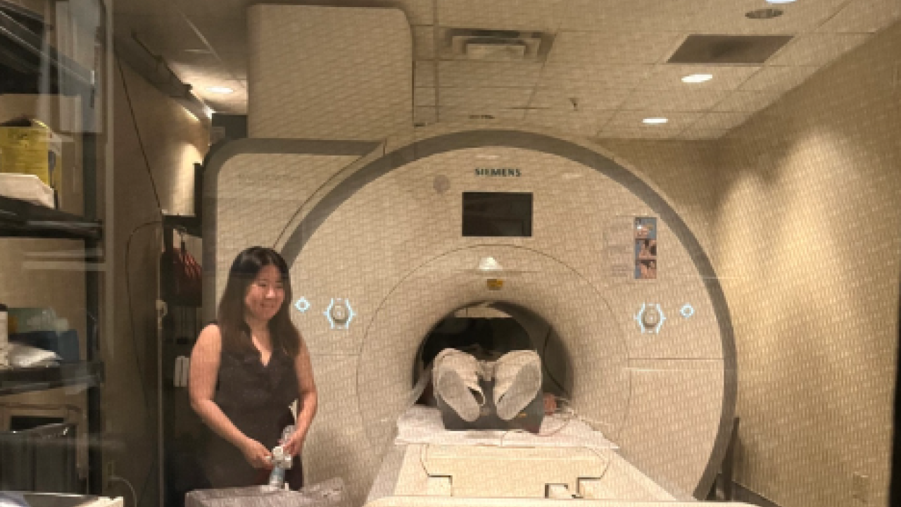
Vascular fingerprinting is a newly developed Magnetic Resonance Imaging, or MRI, technique that compares brain scans against images in a database. Biomedical engineering Ph.D. student Greg Wheeler, in the Fan Lab at the University of California, Davis, leads a project exploring how the method can provide more precise information than traditional MRIs to identify cognitive diseases earlier than ever before.
Building on Existing Technology
MRI is used in clinical settings to take images of tissues inside the body. The patient lies in a tube with a strong magnetic field that aligns the water protons throughout one's body. Radio frequencies are then introduced in a prescribed fashion to stimulate these protons and their specific responses is what shows up in the images.
Traditional MRI generally takes 30 to 60 minutes to generate several scans. These scans are typically qualitative, meaning physicians compare them against images of healthy tissue to detect abnormalities.
Vascular fingerprinting, however, focuses on the brain's vascular system, including blood flow and vessels. This type of scan is quantitative, collecting numerical measurements of the organ that can be precisely compared against other people and over time. It's also much faster, taking about five minutes to produce multiple images.
"If I have quantitative numbers," Wheeler said, "I can see exactly how things changed over six months, whereas in these qualitative images of traditional MRI, it's more difficult to make this kind of precise, long-term assessment."
Identifying Matches
When scanned, a human fingerprint gets compared to a database to find a match. Similarly, a vascular fingerprint MRI is compared to a database of computer-simulated images until the best match is found.
The computer-simulated images are developed by making a biophysical simulation at the same time as the MRI scan. The simulations involve thousands of virtual brain tissues with varying vascular components, such as blood volumes and oxygen saturations. Researchers can compare the simulated images with the original MRI to find the simulated image that most closely matches the real one. Once this match is made, researchers can derive quantitative information from the known parameters — i.e., those vascular components — in the simulated image.
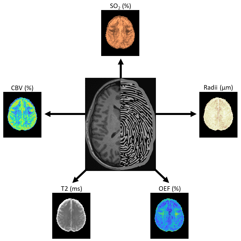
"You get unique 'fingerprints' in your database. You reference that to your acquired image, and you can get these multiple vascular factors from a single scan," said Wheeler.
Currently, Wheeler is studying vascular fingerprinting to develop tools to measure vascular function changes. His work is based on the idea that vascular disease may underpin cognitive disorders like Alzheimer's disease.
"Some of these vascular factors are treatable and are potentially preventable," he said. "If we saw these functional vascular biomarkers before someone developed cognitive impairment, maybe a doctor can prescribe them something to lower their cholesterol or change their diet."
While this technology is only in its beginning stages, Wheeler believes this research is a crucial step in establishing similar tools for doctors to find solutions to these difficult diseases.


