Ph.D. Student Earns Prestigious Early Career Researcher Award from Physics in Medicine & Biology
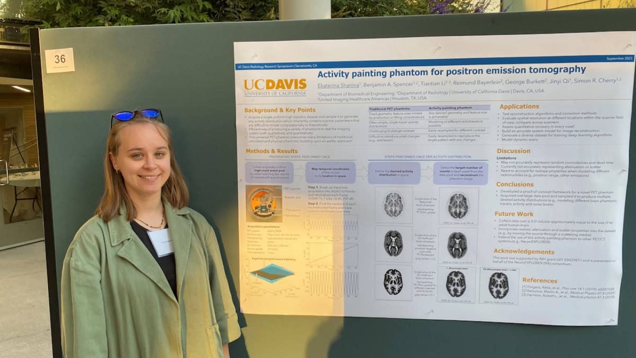
The honor recognizes Ekaterina Shanina's study of a unique PET phantom called PICASSO
Ekaterina Shanina, a Ph.D. student in the Department of Biomedical Engineering at the University of California, Davis, has won the Physics in Medicine & Biology Early Career Researcher Award for her research paper describing a novel brain phantom for positron emission tomography (PET).
Shanina’s study was chosen by Physics in Medicine & Biology’s editorial board as the “best paper” (based on the quality of scientific content and peer review ratings) in the journal’s Early Career Researcher Focus Collection 2024 – a program established to support and highlight the work of emerging researchers in the medical physics and biomedical engineering community.
“The initiative recognizes that early-career researchers often produce cutting-edge, high-impact work but may not yet have widespread visibility,” says Emma Harris, a guest editor on the collection. She explains that while the collection itself showcases a broad range of high-quality work, the award was introduced to further recognize an outstanding contribution from an early-career author – defined this year as someone who completed their Ph.D. in 2018 or later.
“The award serves to highlight exceptional research that stands out for originality, rigor or impact,” says Harris, from the UK’s Institute of Cancer Research and Royal Marsden NHS Trust. “[It will] promote prestige and visibility to the awardee within the international research community, and provide a tangible form of encouragement and recognition that can support academic career progression.”
A new phantom for high-performance PET
In her award-winning paper, PICASSO: a universal brain phantom for positron emission tomography based on the activity painting technique, Shanina describes a unique PET phantom called PICASSO and shows how it can be used to model realistic static and dynamic neuroimaging PET studies with excellent quantitative accuracy.
PET imaging offers an invaluable tool for studying the brain, prompting recent interest in developing advanced high-resolution PET scanners dedicated to brain imaging. Such developments create an associated requirement for appropriate imaging phantoms to evaluate and optimize scanner performance. The PICASSO phantom aims to meet these needs.
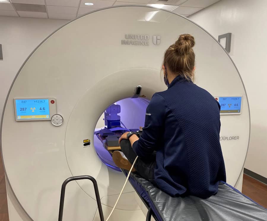
“UC Davis has been collaborating with Yale University and United Imaging Healthcare to develop a new high-performance brain PET scanner called the NeuroEXPLORER,” Shanina explains. “This scanner has high spatial resolution, which renders the most commonly used anthropomorphic brain phantom – the Hoffman phantom – unsuitable for evaluating its performance. At the same time, we wanted to explore the activity painting technique to create this unconventional phantom for PET imaging.”
Most physical PET phantoms need to be filled with a radioactive solution, which means that they can only model one type of tracer and making changes to the phantom structure is challenging. Such phantoms also require walls to separate different regions, which interferes with quantitative image evaluation, and designs with complex internal cavities are hard to fill without residual air bubbles.
“Our PICASSO phantom overcomes many of these limitations,” says Shanina.
It works by moving a 22Na point source around within the field-of-view of a PET scanner to “paint” one high-statistics dataset. The motion of the radioactive source is controlled by a robotic arm and contrast levels are defined by computationally sampling the acquired dataset. This approach can efficiently generate phantoms with arbitrary static and dynamic activity distributions in the brain (or other body regions) using a single PET acquisition.
“PICASSO uses a single dataset acquired with a sealed point source to efficiently generate a variety of activity distributions of various complexities and with arbitrarily fine features,” Shanina explains. “There’s no need for cumbersome phantom preparation, there are no cold walls or air bubbles, and the data contain some of the scanner parameters that are difficult to model analytically. We can even use it to model dynamic studies, which is a very challenging task for conventional phantoms.”
Since the paper was published last year, Shanina and colleagues have extended the two-dimensional PICASSO phantom into a 3D version that can generate whole-brain images. “We are also working on an exciting new application for the phantom, using it to model different time-of-flight resolutions of PET scanners,” she says. “To our knowledge, you cannot do this with any other phantoms that are not simulations.”
Shanina tells Physics World that she is “honored and humbled” to win the Early Career Researcher Award.
“I am very happy that this work keeps attracting people’s attention and interest,” she says. “Of course, I don’t do this all by myself. I am very grateful to have [Distinguished Professor Emeritus of Biomedical Engineering] Simon Cherry and [Professor of Biomedical Engineering] Jinyi Qi as my advisors supporting and encouraging me on this journey.”