New cardiovascular imaging approach provides a better view of dangerous plaques
Combining FLIM with polarization sensitive OCT could lead to new ways to prevent heart attacks and strokes
Researchers in the Department of Biomedical Engineering at the University of California, Davis, have developed a new catheter-based device that combines two powerful optical techniques to image the dangerous plaques that can build up inside the arteries that supply blood to the heart. By providing new details about plaque, the device could help clinicians and researchers improve treatments for preventing heart attacks and strokes.
Atherosclerosis occurs when fats, cholesterol and other substances accumulate on the artery walls, which can cause these vessels to become thick and stiff. A heart attack or stroke may occur if an atherosclerotic plaque inside the blood vessels ruptures or parts of it break off.
“Atherosclerosis, leading to heart attacks and strokes, is the number one cause of death in Western societies — exceeding all combined cancer types — and, therefore, a major public health issue,” said Laura Marcu, research team member leader and professor of biomedical engineering at UC Davis. “Better clinical management made possible by advanced intravascular imaging tools will benefit patients by providing more accurate information to help cardiologists tailor treatment or by supporting the development of new therapies.”
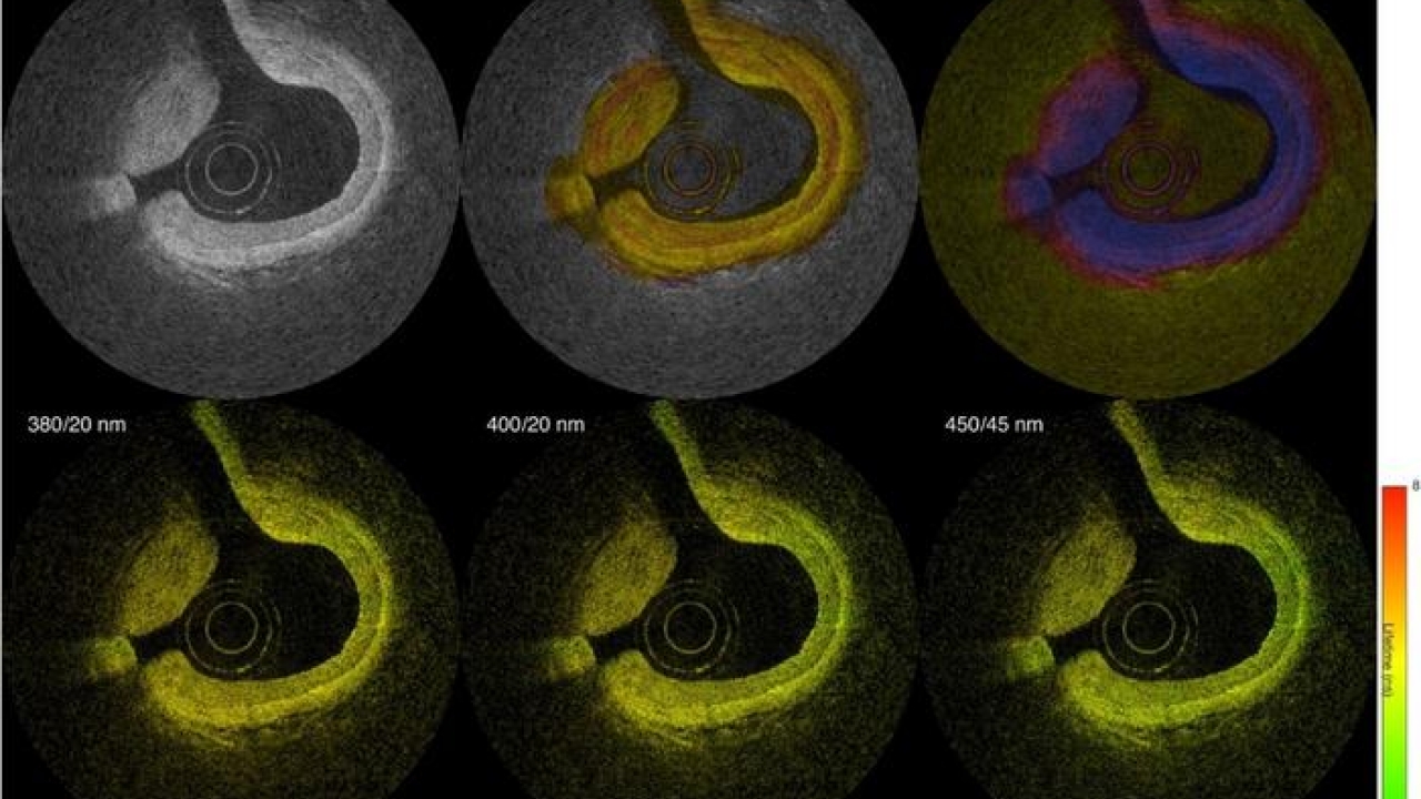
In the Optica Publishing Group journal Biomedical Optics Express, researchers describe their new flexible device, which combines fluorescence lifetime imaging (FLIM) and polarization-sensitive optical coherence tomography (PSOCT) to capture rich information about the composition, morphology and microstructure of atherosclerotic plaques. The work was a collaborative project with Brett Bouma and Martin Villiger, experts in OCT from the Wellman Center for Photomedicine at Massachusetts General Hospital.
“With further testing and development, our device could be used for longitudinal studies where intravascular imaging is obtained from the same patients at different timepoints, providing a picture of plaque evolution or response to therapeutic interventions,” said Julien Bec, first author of the paper and engineering and operations director in the Marcu Laboratory. “This will be very valuable to better understand disease evolution, evaluate the efficacy of new drugs and treatments and guide stenting procedures used to restore normal blood flow.”


