Seeing Light in 'Impossible' Places
New two-photon microscopy system provides more than sixtyfold signal increase over conventional techniques
Researchers at the University of California, Davis, have developed a fast and cost-effective two-photon microscopy system capable of imaging depths previously impossible to reach in scattering tissues, such as bone and the brain.
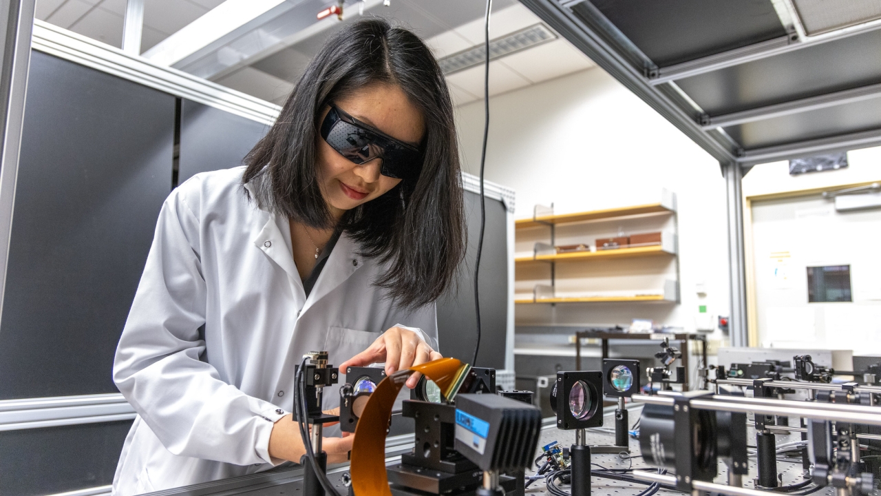
Led by Assistant Professor of Biomedical Engineering Yi Xue, the system shows immense promise to enable scientific insights into biological features once only observable after surgical removal, like the anatomy of neurons deep in the brain. This is because the system boosts the signal strength of deep tissue microscopy systems by over 60 times while being faster and cheaper to produce than similar microscopy systems.
"We want to visualize things previously invisible through traditional deep tissue imaging methods," Xue said.
Xue and her team described their new system, 2P-FOCUS, in a paper published in Optica’s Photonics Research Journal. The research is the direct result of Xue’s Maximizing Investigators' Research Award from the National Institutes of Health, awarded in 2024.
Mirror Image
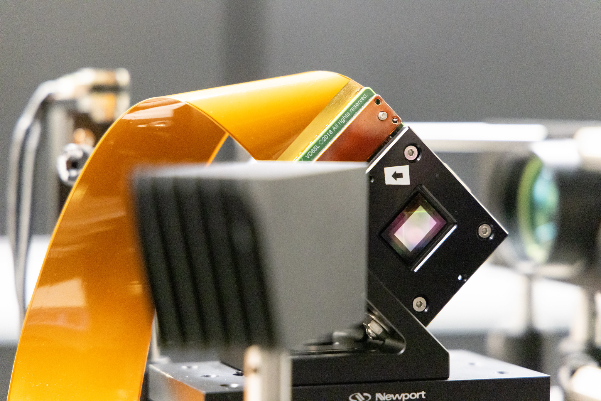
2P-FOCUS uses two-photon excitation microscopy, an existing technique for imaging living tissue by shining light on molecules that can radiate light back. The new system improves upon this technique with an innovative method for correcting light scattering, thanks to a component commonly found in classroom projectors: a digital micromirror device.
Digital micromirror devices, or DMDs, manage the flow of light with millions of mirrors, each about 10 micrometers big — over 20 times smaller than a penny. Each mirror can be programmed to flip between two positions: one to block light and one to allow light to pass through at particular points, such as a single pixel of an image.
In the case of 2P-FOCUS, a DMD allows researchers to use light more efficiently by marking the exact areas where light needs to penetrate for deep tissue imaging.
"The standard two-photon microscope shines light on a sample without any selection," Xue said. "Say we have a certain amount, like 10 milliwatts of light, shining on a sample. Nine milliwatts of that light will go to random places, becoming heat energy, and only one milliwatt will work to image what we want to see."
Think of it like a flashlight pointed at a tall picket fence from a few feet away in the dark of night. The light emanates from the flashlight and illuminates the wood panels, with only some light passing through the fence’s gaps. Behind the fence is a large oak tree, but it’s hard to make it out due to the light barely hitting it.
If a DMD system were applied in this scenario, the flashlight user could focus all of the light to fit through the gaps of the fence and beam directly onto the tree, revealing the deep and rough fissures of the oak tree’s bark with clarity.
Light Work
For Xue’s team to see the brain structures hidden deep beneath the skull — aka, their oak tree behind the fence — they have to run hundreds of experiments. These experiments randomly shine light across their sample to see where light can reach deep into the recesses of the brain and where it is hitting the outermost layer.
This is tracked through the tissue’s strength of fluorescence, or the brilliance of reflected light, under illumination. Areas that excite with high fluorescence show as white or near-white — these are where the light is able to probe the scattering tissue — and areas that are dark grey to black are where the light is dispersing into heat energy.

They map this data to a grid corresponding to the pixel-like layout of the DMD. Xue and her team then use this grid to configure 2P-FOCUS to control the flow of light, guiding it to the areas where it was observed to probe deep into brain tissue.
It is through this high-power, sharply focused light that 2P-FOCUS is able to image tissues found deeper in the body than other two-photon microscopy systems. The resulting images are road maps of tissue and can show fine details like the neuronal structure of the brain.
Beyond the Depth of Field
The DMD component in Xue’s 2P-FOCUS advances more than the imaging depth for deep tissue imaging. It also increases the speed of two-photon microscopes with scattering correction and greatly lowers their cost when compared to other systems.
Traditional two-photon microscopes that have the capability of scattering correction use spatial light modulators. These rely on liquid crystals to control the flow of light. Top-of-the-line spatial light modulators for microscopes can cost upward of $20,000 and reach a peak refresh rate of 60-240 hertz — essentially, the speed at which the device can control light.
A high-end DMD outperforms both of these metrics.
"For a few thousand dollars, a DMD can go over 10 kilohertz easily," Xue said. "A very good one can even reach 100 kilohertz. So, our system is about 100 to 1000 times faster than those with spatial light modulators and at a fraction of the cost."
Imaging the ‘Impossible’
The 2P-FOCUS system can be broadly applied to improve deep tissue imaging. Or in Xue’s words, to see the unthinkable.
"With 2P-FOUCS, we can see things people previously thought were impossible to see."
One potential application of the microscopy system is to improve researchers' ability to probe and understand the mechanics of neurodegenerative diseases, such as Alzheimer’s.
Xue is most interested in using it to understand how people actively learn information and consolidate memories through deep tissue imaging of the hippocampus, where learning and memory consolidation occur.
"All these kinds of processes currently cannot be studied using traditional optical methods in the living animal, but our technique may allow researchers to do so in the future."
"That’s the big picture for us," Xue said. "Our next step is to go even deeper than what we’ve shown in our Optica Photonics Research paper to reach important brain regions previously untouched by deep tissue imaging. Through these future studies, we hope to learn very complex and large-scale mechanisms of the brain that underlie significant behaviors, such as memory formation."