


Improving Cancer Treatment with Digital Twins
Imagine a doctor being able to individualize treatment for liver cancer with a digital replica of their patient’s body. Such a tool would ensure greater success rates and improved safety in treating one of the world’s deadliest cancers.

That’s the future envisioned by Emilie Roncali, an associate professor of biomedical engineering at the University of California, Davis. In new research supported by funding from the National Cancer Institute, she’s developing a digital twin for the precise care of liver cancer.
Her digital twin will allow physicians to plan, on a patient-to-patient basis, for a new liver cancer treatment technique called yttrium-90 radioembolization, or 90Y TARE. Unlike traditional chemotherapy’s generalized radiation, the technique targets tumors and is emerging as one of the most effective interventions for liver cancer.
However, a large number of patients who receive 90Y TARE are currently underdosed due to fear that the radiation will cause liver toxicity, leading to unsuccessful treatments.
“This problem is due to the lack of precise tools to plan the treatment,” Roncali said. “If we had models to predict the radiation dose before the treatment, patients could receive a much more efficient therapy with a higher radiation dose precisely targeting the tumor.”
False Mirrors
The reality of healthcare is that each patient is slightly different, with no two patients mirroring one another. These individual differences are small and often unremarkable, but add up when a treatment’s success operates at the margins of the microscale.
In the case of liver cancer, it’s the minute variances in the vascular system, or the network of vessels carrying blood throughout the body.
“The vascular system is amazingly similar in all human beings, but not completely the same,” Roncali said. “That is why this treatment is so difficult to plan, because the patients need to go for that exploratory workup.”
This is because the liver receives blood supply from two sources. The first is the portal vein, which supplies about 75% of blood to the organ. The other is the hepatic artery system, which supplies the remaining 25% to healthy liver tissue, but close to 100% of the blood feeding cancerous tumors.

The tumors’ bias toward the hepatic artery system is what 90Y TARE exploits. Doctors can inject radioactive particles — a few tenths of a millimeter large — into the bloodstream to target each tumor. The issue is that current imaging techniques struggle to capture the differences between each patient’s hepatic artery system, leading to cautious radiation doses without reliable guidance.
Precision from Personalization
Roncali’s digital twin will capture the unique features of each patient’s vascular system and allow doctors to predict blood flow and how the radioactive isotopes will break down in the body.
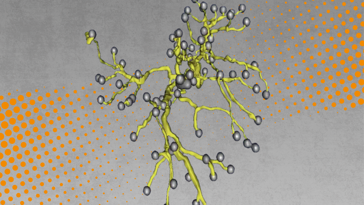
The twin is created by pulling patient data from several biomedical imaging techniques, such as CT scans. Roncali’s team couples these images with a mathematical model called CFDose to create a three-dimensional, individualized digital model of the patient’s vascular system and liver, allowing doctors to visualize distinctive branching paths of the vasculature and simulate the impacts of different doses of 90Y Tare.
CFDose is a predictive modeling technique that Roncali innovated in 2020, notable for its ability to combine computational fluid dynamics (think: how blood moves) with radiation physics modeling. These dual components enable the digital twin to predict both the movement of radioactive microspheres in a patient’s bloodstream and the spread of radiation throughout the body, ultimately creating free radicals — the unstable molecules that kill cancer cells.
“The system is really good at predicting how blood flow will move through the volume, through the arterial tree forest,” Roncali said. “The role of the twin is to take that exact information unique to the patient and run a model to predict where those microspheres will go to deliver the radiation.”
Through these capabilities of the digital twin, physicians will be able to plan precise and personalized dosages of 90Y TARE for the treatment of liver cancer.
A Living Document

A description of a digital twin could be this: A one-to-one virtual representation of a physical system used for conducting tests or predictions. What, then, makes it better than a computational model in a medical context?
“Both aim to represent the patient as best as possible, but the digital twin should go both ways. It should inform how we treat or monitor the patient and be updated in real time,” Roncali said.
This bi-directional, real-time relationship between the physical and virtual defines a digital twin. If the twin was static, like a computational model, it wouldn’t allow physicians to continue providing precise care.
“You're monitoring the therapy, you're updating the therapy, saying, ‘Okay, the chemotherapy or radiation is going there. It's not good. We need another treatment,’” Roncali said. “Imaging guides your therapy very precisely, and then you can see how the treatment works with the same model to inform future therapeutic interventions. It’s like a feedback loop.”
Model to Follow
Roncali’s work to improve the treatment outcomes of liver cancer is part of a larger effort to advance human health with digital twins.
“There's a lot of work on digital twins right now. I think the next challenge will be, if we had a twin of the whole human body, could we have a connection between organs?”
In the case of Roncali’s model, she’s already thinking about ways to use her digital twin as a model for future advancements in health care, adapting it for different pathologies or parts of the body.
Her recently elected position as director of biocomputational medicine for the Center for Precision Medicine and Data Sciences at UC Davis Health will supercharge this effort. There, she will set the stage for what’s possible with digital twins.
“In the near future, I don’t see a digital twin as an avatar of our body, but rather as a tool dedicated to a clinical task — cancer-related or not — to facilitate the medical process and improve the patient’s outcome.”


